Showing posts with label cancer. Show all posts

cancer lungs: 14 PREVENTION


The most important prevention is not smoking from an early age. Quitting smoking can reduce the risk of lung cancer. Research from the group of smokers trying to quit smoking, only 30% were successful.

Lately a lot of preventive chemopreventif done. Aims to intervene Chemopreventif carcinogenic process, using natural or pharmacological drugs. Kemoprevensi agents are widely used micronutrients or antioxidants, retinoids, carotenoids, vitamin C, selenium, hormone therapy. Chemopreventif agents must be accompanied by experimental or epidemiological data showing efficacy, the mechanistic basis for the activity and same granting chemopreventif the long term.

Conclusion:
1. In men, lung cancer and the leading cause of death in women, lung cancer the third leading cause of death.
2. Practical division for medicinal purposes: a). small cell lung cancer (SCLC), b). NSCLC (non small cell lung cancer / squamous carcinoma, adenocarcinoma, large cell carcinoma).
3. Treatment for lung cancer depends on the type, the SCLC is chemotherapy treatment, radiotherapy for NSCLC, while surgery is the best option.
Read More - cancer lungs: 14 PREVENTION

cancer lungs: 13 THERAPY


Treatment of lung cancer, depending on the type and stage of cancer, in addition to the overall health of patients. If the patient has emphysema, for example, the poor pulmonary function inhibits the operation on patients, even if the patients have tumors that can be surgically removed. Other factors may also play a role, no matter what type of cancer patients. There was a time where, for example, when the side effects of treatment greater than the benefits gained. If that happens, patients can be given supportive therapy. This means, treat symptoms of cancer, causing pain and difficulty in breathing, but does not cure the cancer itself.

The purpose of lung cancer treatment are:
 Curative: cure or prolong disease-free period and increase the life expectancy of patients.
 Palliative: reducing the impact of cancer, improve the quality of life.
 Treat home (Hospice Care) in the case of terminals; reduce physical and psychological impact of cancer both in patients and families.
 Supportive: supporting palliative and curative treatment such as terminal nutrition, blood transfusion and blood components, growth factor anti-pain medication and anti-infective drugs.

There is a fundamental difference of biological temperament Small Cell Lung Cancer in Non-Small Cell Lung Cancer, so the treatment must be distinguished:

Small Cell Lung Cancer (SCLC)

Because most small cell lung cancer has spread outside the lungs when found, surgery is usually not an option. The most effective treatment is chemotherapy, either as monotherapy or in combination with radiation therapy.

SCLC is divided into two, namely:
1. Limited stage disease who were treated with curative purposes (the combination of chemotherapy and radiation) and treatment success rates of 20 %.
2. Extensive-stage disease treated with chemotherapy and initial treatment response rates of 60-70% and complete treatment response rates for 20 - 30 %. Median survival time for limited disease state is for 18 months and extensive disease state is 9 months.

Radiotherapy. In some cases inoperable, radio therapy performed as curative and can pengobatab as adjuvant therapy / palliative in tumors with complications are like reducing the effects of obstruction / suppression of blood vessels / bronchus.

This therapy uses X-rays to kill cancer cells. In some cases, radiation may come from outside the body (external radiation). On the other hand, radioactive compounds can be placed on the needle, using a catheter inserted into or near the lung (internal radiation). How radiation given depending on the type and stage of cancer is handled. Radiation therapy can be administered before, during, or after chemotherapy. In all cases, the goal of treatment is to destroy cancer cells with as little as possible interfere with the normal tissue.

Medication side effects may include redness and swelling of the skin, where the radiation enters the body, shortness of breath, fatique, and sometimes hard to swallow. Dysphagia due to post radiation esophagitis often occurs while the post radiation pneumonitis are rare ( <> 50 % tumor measured 50 or more than 5 the number of detected lesions disappeared; c). stable disease 50 % reduction or <> 25 % bigger; e). lokoprogresif: tumors growing within a radius of the tumor (local).

Side effects of chemotherapy is the most disturbing aspect of the treatment of cancer cells with rapid growth. As contained in the digestive tract, bone marrow and hair, is part of the most influential of these drugs. Although many side effects occurred, severity of cancer depends on these drugs. Sometimes patients have several reactions. On the other hand, patients may experience symptoms such as nausea and vomiting, dizziness, feeling very tired and the risk of infection increases.

Non-Small Cell Carcinoma
Surgical therapy is the first choice in stage I or II in patients with adequate reserves remaining lung. In stage IIIA there is still controversy about the success of the operation if the ipsilateral mediastinal lymph or there thorax wall metastases. Tumor removal technique performed a variety of techniques. Thoracotomy or the opening of the chest wall for surgical procedures and operations sternotony median or do by cutting through the breastbone is the standard method for lung cancer surgery.

Operation to treat lung cancer include:
 Wedge resection. In this operation carried out removal of the lung tumors, along with the soft tissue margin.
 Lobectomy. Operation of lung cancer most often committed. Lobectomy is the appointment of the entire lobe of one lung.
 Pneumonectomy. In this operation, the entire lung removed. Because pneumonectomy would reduce lung function, and cause other complications, this action is only done when necessary and if the patient is able to breathe with one lung.

Operating procedures will also have side effects that can cause lymphocytopenia or low number of lymphocytes (white blood cells) in the blood that causes the short survival time in patients with advanced-stage cancer.

The use of chemotherapy in NSCLC patients in the last two decades has been investigated. Curative chemotherapy for the treatment of combined integrated with other cancer treatment modalities in patients with advanced disease lokoregional.

Chemotherapy is used as standard therapy for patients ranging from stage IIIA and for palliative treatment. Sitostatika drugs have good activity in NSCLC with response rates between 15 - 35 %, however the use of a single drug did not achieve complete remission.

Combination has been investigated sitostatika to increase response rates that will have an impact on life expectancy. According to the Food and Drug Administration (FDA), the use of Avastin with paclitaxel and carboplatin can be used for initial systemic therapy in patients who can not do surgery. For patients with metastatic lung tumors in the colon and rectum, Avastin can be used as sitostatika combined with intravenous 5 - fluorouracil.
Read More - cancer lungs: 13 THERAPY

cancer lungs: 12 STAGING


Staging is a system of classifying information about cancer, including location and how far the cancer has spread. TNM system is often used in establishing the stages of lung cancer, especially for Non-Small Cell Lung Cancer. Lung tumors are classified according to tumor size (T), the level of lymph node involvement (N), and how far the cancer has spread (M). Staging Non-Small Cell Lung Cancer include:
 Stage 0. At this stage, limited cancer of the lining of the airways and lung tissue had invaded. 0 cancer, is often found during bronchoscopy performed, which may be done to see the X-ray abnormalities in the chest. When found and treated as soon as possible, of cancer at this stage can be eliminated.
 Stage I. Cancer at this stage has invaded lung tissue, but has not spread to the lymph node.
 Stage II. At this stage the cancer has spread to surrounding lymph nodes and invade the chest wall
 Stage IIIA. At this stage, the cancer has spread from the lungs to the lymph node in the center of the chest.
 Stage IIIB. The cancer has spread to areas such as the heart, blood vessels, trachea and esophagus or to the lymph nodules in the collarbone area.
 Stage IV. The cancer has spread to other parts of the body such as the liver, bones or brain.

Staging for Small Cell Lung Cancer is different from Non-Small Cell is divided into limited and extensive:
 Limited. Cancer of the lungs and nearby lymph nodes.

 Extensive. The cancer has spread outside the lung and surrounding lymph nodes, and may have attacked the lungs or other organs.

Staging test used to determine how far the cancer has spread is important in planning treatment. In addition to CT scan, this test includes:
 Magnetic Resonance Imaging (MRI). In addition to radiation, this test uses radio waves and powerful magnets to produce images of body parts. This is very good for detecting tumors that have spread to the brain or spinal cord.
 Positron Emission Tomography (PET) scans. Unlike other imaging techniques, PET scans do not produce images clear organ structure. Even this way of producing color images of the area with more or less intense to provide information about chemical activity within certain organs and tissues. This chemical activity may indicate whether the cancer cells have spread to nearby lymph node, even before the enlarged lymph node. However, PET scans should be interpreted carefully, because sometimes benign conditions can mimic cancer.
Read More - cancer lungs: 12 STAGING

cancer lungs: 11 Prognosis


A. Small Cell Lung Cancer (SCLC):
 With a change therapy in the past 15 - 20 years the possibility of life on average (median survival time) who had < 3 months increased to 1 year.
 The possibility Disease Limited group average life up to 1-2 years, while 20% of it remained alive in 2 years.
 30 % died because of local complications of the tumor.
 70 % died of carsinomatosis.
 50 % metastasize to the brain (autopsy)

B. Non-Small Cell Lung Cancer (NSCLC):
 Most important in the prognosis of lung cancer is to determine the stage of the disease.
 Compared with other types of NSCLC, karsinomaskuamosa not as bad as others. In patients who performed surgery, the possibility of life after 5 years was 30 % surgey.  Survival after surgery, 70 % on Occult carcinoma; 35 - 40 % in stage 1; 10 - 15% in stage II and less than 10 % at stage III.
 75 % of squamous carcinoma died of complications torakal, 25 % because of the extra torakal, 2 % of them died because of the central nervous system disorders.
 40 % of adenocarcinoma and large cell carcinoma died of complications torakal, 5 for 55 extra torakal.
 15 % of adenocarcinoma and large cell carcinoma metastatic to the brain and 8 - 9 % die from central nervous system disorders.
 The possibility of life on average tumor metastases patients varies from 6 months to 1 year, where it is highly dependent on performance status (Karnofsky scale), extent of disease, the weight loss in the last 6 months.
Read More - cancer lungs: 11 Prognosis

cancer lungs: 10 DIAGNOSTIC PROCEDURES

A. Photo chest roentgens in Posterior-anterior (PA) and lateral
Simple initial examinations that can detect lung cancer. Mayo Clinic Study of the USA, found 61 % of lung tumors were detected in routine examination with normal chest roentgens images, whereas sputum cytologic examination can detect only 19 % . In lung cancer, breast examination roentgens photo also necessary to re-evaluate its doubling time. Reported that most lung cancer has a doubling time of 37 - 465 days. When the doubling time> 18 months, mean tumor benign. The signs of other tumors are benign lesions, concentric spherical, solid and calcification of the firm.

Inspection photos chest roentgens tomography more accurate ways to support the possibility of lung tumors, when the usual way breast images can not confirm the presence of tumors. Investigations another radiologist who sometimes is also necessary bronkografi, fluoroscopy, superior vena cavografi, ventilation / perfusion scanning, ultrasound sonography.

In photos roentgens primary lung cancer can be seen as a solitary round shadow or cause the collapse of a lobe due to bronchial tumor block. Early cancer can be very small and irregular shape, but usually will be fine when growing up. While the picture radiologist for secondary lung tumors does not indicate where the location of the primary tumor.

B. Examination Computed Tomography and Magnetic Resonance Imaging

CT scan at thorax, more sensitive than normal chest image examination, because it can detect abnormalities or nodules with a minimum diameter of 3 mm, although false positives for abnormalities of this magnitude can reach 25 - 60 %. If this facility possible, a CT scan could be a second screening examination after normal chest images. Magnetic Resonance Imaging examination (MRI) is not routinely done, because he is limited to assessing the invading tumor abnormalities in the vertebrae, spinal medulla, the mediastinum, in addition to cost is also quite expensive.

MR examination thorax not superior to CT scan thorax. We are developing a technique that is more accurate imaging of Positron Emission Tomography (PET) can distinguish benign and malignant tumors based on differences in metabolism of biochemical substances case of glucose, oxygen, proteins, nucleic acids. Examples of substances used methionine 11 C and F - 18 fluorodeoxyglucose (FD6).

Tumors less than 1 cm, somewhat difficult to detect because of the small size is less resolusion by PET Scanner. Sensitivity and specificity reported how PET is 83-93% 60-90% sensitive and specific.

Some false positives to be found also a sign of malignant lesions such as inflammation and infection and tuberculosis aspergilosis. Although it is known from several studies PET examination has better accuracy values than CT scans.

C. Inspection of Bone Scanning

This examination is required if the expected signs of metastases to the bone. Tumor incidence of non small cell lung cancer (NSCLC) to bone was reported by 15 %.

D. Sputum cytology examination

Sputum cytology examination is done routinely, especially when patients have complaints such as cough. Cytologic examination does not always give positive results because it depends on:
 Location of tumors of the bronchus
 Type of tumor
 Engineering issued a sputum
 The number of sputum examination. Examination is recommended 3 - 5 days in a row
 When sputum examination (sputum must be fresh)

In lung cancer, which is central to good sputum examination can give positive results up to 67 - 85 % in squamous cell carcinoma. Sputum cytology examination is recommended as a routine examination and screening for early diagnosis of lung cancer, and they are developing early diagnosis using sputum examination staining with immune MAB with antigen antibody 624 H 12 for SCLC (small cell lung cancer) and 703 D4 antibodies to antigens NSCLC (non small cell lung carcinoma). Reports from the National Cancer Institute USA this technique gives results 91 %, 88 % sensitive and specific.

Other cytologic examination for lung cancer diagnostics can be performed on pleural fluid, aspiration of cervical lymph nodes, supraklavikula, rinse and bronchus on bronchoscopy sweep.

E. Histopathology examination

Histopathologic examination is the gold standard for diagnosis of lung cancer to obtain biopsy specimens in a way through:

Bronchoscopy. Modification of fiber-optic bronchoscopy may include:
 Trans bronchial lung biopsy (TBLB) with the demands of fluoroscopy or ultrasound.
 Recently fuorescence examination was developed by using fluorescence bronchoscopy exchanging agent such as Hp D (hemato porphyrin derivate) gave fluorescence concentrates in cancer tissue. The newer technique is to auto fluorescence bronchoscopy. The results of this examination showed 50% more sensitive than white light bronchoscopy for detection of carcinoma in situ and severe dysplasia.
 Ultrasound bronchoscopy, also developed at this time to detect peripheral tumors, endobronkial tumors, lymph node lesions of the mediastinum and hilar regions.
 positive result with bronchoscopy can be reached: 95 % for centrally located tumors and 70-80% for tumors of peripheral location.
 Trans-bronchial-Needle Aspiration (TBNA). Working against lymph nodules in the hilar or mediastinal. The result will be better when guided by CT scan.

How to examination by bronchoscopy is to insert a tube pipe flexible bronchoscope into the airways. In this way doctors can look inside the lungs and lung tissue samples taken for laboratory examination.


Trans Torakal biopsy (TTB)
If the patient has cavities fluid in the lungs, the doctor may take a sample by inserting a thin needle into the chest between the ribs. Fluid is then examined in the laboratory, to see whether or not cancer cells. Raised a number of liquids with a biopsy or torasentesis can also improve breathing.

Biopsy with TTB especially for lesions located peripheral to the size of < 2 cm sensitivity reached 90 - 95 %. Complications pneumothorak can reach 20 - 25 % and haemoptysis up to 20 %. With better preparation, these complications can be minimized. Examination results will be better if there is demand for CT scans, ultrasound, or fluoroscopy. Biopsy of lymph nodes palpable, can be done in Daniel's biopsy of the lymph-nodes scalaneus supraklavikular. Torakoskopi. Biopsy of the tumor in the pleura give better results than by way torakoskopi blind (blind). For tumors located on the surface of the pleural biopsy with the way visceralis Video Assisted Thoracoscopy has a sensitivity and specificity to 100 %, whereas complications occur is very small. Mediatinoskopi. More than 20 % of lung cancers metastasize to the mediastinum, especially Small Cell and Large Cell Ca Ca. to obtain tumor metastases or lymph nodes involved can be done by mediastinoskopi where mediastinoskopi inserted through the supra-sternal incision. This can help assess how far the cancer has spread and whether the operation is wise choice to remove the tumor. Biopsy results positive value 40 %. From other studies on the false negative values obtained mediastinoskopi of 8 - 12 (followed by Thoracotomy).

Thoracotomy for lung cancer diagnostic procedures done when non-invasive and invasive previously failed to find tumor cells.
Read More - cancer lungs: 10 DIAGNOSTIC PROCEDURES

cancer lungs: 5


Like most other cancers than the exact cause of lung cancer are unknown, but prolonged exposure or inhalation of a carcinogenic substance which is the main causative factor in addition to other factors such as immune, genetic, and others.

From some literature has reported that the etiology of lung cancer is related to smoking. Lombard and Doering (1928) have reported high incidence of lung cancer in smokers compared with nonsmokers.

Layer formed from the respiratory tract cells to the surface of a rectangular shape (columnar epithelium) and the glands that produce mucus and other fluids. In the lungs of healthy, these cells divide in a controlled and orderly arrangement. However, when a cell becomes cancerous, the cells continued to divide even when new cells are not needed. Although it took years to develop lung cancer, lung tissue changes may occur as soon as possible after exposure with lung cancer causing compounds (carcinogens) in cigarette smoke. When exposed continuously, normal cells are damaged and eventually become cancerous. Because of easy access to the bloodstream and lymph vessels, cancer cells can spread to other parts of the body before the patient develops symptoms.

Smoking is the main cause of approximately 90% of cases of lung cancer in men and about 70% in women. The more cigarettes smoked, the greater the risk for suffering from lung cancer.

There is a relationship between the average number of cigarettes smoked per day with a high incidence of lung cancer. It is said that, 1 of 9 heavy smokers will suffer from lung cancer. Later the report several studies suggest that passive smokers would be at risk for lung cancer. Children who are exposed to smoke for 25 years in adulthood will be subject to the risk of lung cancer doubled compared with the not exposed, and women living with husband / partner is also affected smokers lung cancer risk 2-3 fold. An estimated 25% of lung cancer than non-smokers is derived from passive smokers. Lung cancer incidence in women in the USA in the last 10 years also increased to 5% per year, partly because of the increasing number of women smokers or passive smokers.

Effect of cigarettes not only lead to lung cancer, but can also cause cancer in other organs such as the mouth, larynx and esophagus.

Report of the NCI (National Cancer Institute) in the USA in 1992 declared cancer in other organs such as kidneys, urinary vesika, ovary, uterus, colon, rectum, liver, penis and others higher in patients who smoke than non-smoking.
Read More - cancer lungs: 5

cancer lungs: 4 Mesotelioma Malignant


Bronchial adenoma is a small group of malignant neoplasms of low the agresifitas. Occur in the lower trachea or main bronchi. Two of the most important forms are bronchial carcinoid and rare silindroma. Bronchial carcinoid as small cell carcinoma, derived from cells of bronchial mucosa Kulchitsky. These tumors develop nearly 4% of all bronchial tumors. Can become real in adolescence until middle age (average age at diagnosis, 45 years), where the number of men and women are affected about the same lot. Signs and symptoms of bronchial obstruction such as chronic cough, haemoptysis, or pneumonitis are common. Bronchial carcinoid tumors like carcinoid of the small intestine. Some tumors secrete serotonin, 5-hydroxy tryptophan, and other biologic substances which generate a complex of symptoms known as carcinoid syndrome. The symptoms include a red face, bronchoconstriction, and wheezing, and diarrhea. Carcinoid tumors follow the course of the disease is relatively benign, and surgical resection is usually quite useful, given the survival rate 5 years, exceeding 90% of typical carcinoid.

Mesotelioma malignant tumor that is generally not of the pleura, where the majority of cases associated with exposure to an abscess. This exposure may be brief but usually the time between the exposure and clinical awitan is 25 years old. Malignant Mesotelioma very fierce and the survival of less than 1 year from the time of diagnosis.

Both primary sarcoma of lung and primary malignant melanoma both lungs are rare, but it is a form of lung cancer are very fierce. Usually the type of lung cancer is more of a metastasis from the primary tumor was diagnosed rather than a locus of primary tumors.

Finally, it must be remembered that the lung is more often a place as metastasis cancer than the location of primary malignant neoplasms. The lungs are often the place deposits of cancer cells from the secondary other organs, because microscopic tumor emboli are carried by the blood is usually caught in the capillary network of the lungs. Tumors carried by the lymph from the lower half of the body and the abdominal cavity can be detained while walking through the duct torasikus. Neoplasms that often lead to lung metastases, respectively from the most common carcinoma of the breast, gastrointestinal tract, female genital tract and kidney, melanoma and male genital cancers.
Read More - cancer lungs: 4 Mesotelioma Malignant

cancer lungs: 2


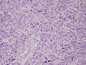
Lung cancer is usually classified according to primary histology types and all have a natural history and response to treatment varies. Although there are more than a dozen types of lung cancer primary, but bronkogenik cancer, including four first types of cells is 95% of all lung cancers.

Practical division for medicinal purposes: a). small cell lung cancer (SCLC), b). NSCLC (non small cell lung cancer / squamous carcinoma, adenocarcinoma, large cell carcinoma).
1999 WHO histological classification for tumors and pleural tumors: (2)
Epithelial tumors:
1. Benign: papilloma, adenoma
2. Preinvasive lesion: squamous dysplasia / carcinoma in situ, atypical adenomatous hyperplasia, diffuse idiopathic pulmonary neuroendocrine cell hyperplasia.
3. Malignant:
1. Squamous cell carcinoma: papillary, clear cell, basaloid.
2. Small cell carcinoma: combined small cell carcinoma
3. Adenocarcinoma:
• Acinar
• Papillary
• bronchoalveolar: nonmusinous, musinous, musinous and nonmucinous mixed or indeterminate cell type

• Solid carcinoma with mucin formation
• Adenocarcinoma with mixed subtypes
4. Large cell carcinoma: neurondocrine Large cell carcinoma, Basaloid carcinoma, Lymphoepitelioma-like carcinoma, Clear cell carcinoma, Large cell carcinoma with rhabdoid phenotype
5. Adenosquamous carcinoma
6. Carcinoma with pleomorphic sarcomatoid or sarcomatous elements
7. Carcicoid tumors: typical carcinoid, atypical carcinoid
8. Salicary gland carcinomas of type: mucoepidermoid carcinoma, adenoid cystic carcinoma
4. Others: soft tissue tumors
5. Mesothelial tumors: Benign, Malignant mesothelioma
6. Miscellaneous tumors
7. Lymphoproliferative diseases
8. Secondary tumors
9. Unclassified tumors
10. Tumor like lesions
Read More - cancer lungs: 2

cancer lungs: 1


Currently, cancer is a major cause of death in the productive age. It is estimated that, at least there are 150 new cancer patients each year per 100,000 people in Indonesia. This means, of the 200 million population is estimated there will be 300,000 cancer patients each year.

The prevalence of lung cancer in developed countries is very high, in the USA in 2002 there were 169,400 reported new cases (of which 13% of all new cancers are diagnosed) with 154,900 deaths (of which 28% of all cancer deaths), the prevalence of events in the UK reached 40,000 / year, while in Indonesia was ranked 4th largest cancer, the cancer hospital Dharmais occupied Jakarta in 1998 ranked third after breast cancer and cervix. The death rate from lung cancer worldwide reach approximately one million residents each year. Because of our recording system that prevalence was not good but certainly not yet known, and pulmonary tumor clinic at the hospital felt the true increase. In other developing countries reported the incident quickly rising partly because of excessive tobacco consumption in China is like 30% of cigarettes consumed the world. Most of the male lung cancer (65%) life-time risk in women 1:13 and 1:20.

Lung cancer is one of the biggest causes of death worldwide, with incidence rates increasing from time to time. Among all the diseases of cancer in men, lung cancer is the most common cause of death in the world. In women although the number
low incidence of lung cancer but it is the third leading cause of death due to cancer.

More than 1.3 million new cases of lung and bronchial cancer worldwide, causing 1.1 million deaths each year. Based on the number of incidents and the prevalence in the world, Asia, Australia, and Far East are at the first level with an estimated number of cases more than 670 thousand with a mortality rate of more than 580 thousand people. In Indonesia, lung cancer became the main cause of death of men and more than 70% of new cancers were diagnosed at an advanced stage (stage IIIB or IV). Therefore, only 5% of patients can survive for up to 5 years after being found positive.

According to Ahmad Hudoyo from Friendship Hospital, in Indonesia, lung cancer is the most cancers. This is mainly because the number of smokers, particularly smokers is very much a beginner. In the United States and European anti-cigarette programs are running and can hit smokers from 70% to 20%, lung cancer rates are still too many.

Until now, lung cancer is still a major problem in medicine. Difficult to detect lung cancer. In the early stages, this cancer has no symptoms, so that when the patient had been diagnosed at an advanced stage. Cancer cells that are not controlled in the lung tissue, making wild production causing that inhibits tumor growth and stop the lung function as they should. The large size of the lungs, causing cancer to grow for years undetected and without symptoms. The disease is only detected after the cancer reaches an advanced stage.
Read More - cancer lungs: 1